Estimated reading time: 8 minutes
Researchers are increasingly using eye trackers to explore changes in pupil size (pupillometry) in order to reveal insights into cognitive processes. EyeLink eye trackers are capable of detecting changes in pupil size of just 0.1% of the pupil diameter, and their high sampling rates allow pupillary responses to be measured with exceptional detail. This blog outlines some of the key considerations for people interested in doing pupillometry research. For a far more detailed consideration of some of the issues outlined below, I encourage readers to check out Sebastiaan Mathot’s excellent papers on the topic (references at the end of the blog).
Trial Durations are Important to Consider in Pupillometry Research
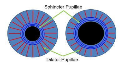
The muscular arrangement that determines pupillary dilation and constriction is well known. Radial muscles dilate the pupil and the circular sphincter muscles constrict it.
The most important point as far as pupillometry research is concerned is that pupil size changes are slow. The muscles involved in both dilation and constriction contain smooth muscle fibers – the same type of muscle fibers that are found in the gut and blood vessels. They excel at making repeated movements with low energy requirements, and do not fatigue like skeletal muscles – but they are slow-moving. Even the fastest movement (constriction in response to bright light) takes place over hundreds of milliseconds. This means that for pupillometry research you need to allow enough time in each trial for any pupillary response to unfold. It also means that a pupillary response on trial N may potentially “spill over” into trial N+1. It is easy to imagine situations in which the impact (for example of a very distressing image) has consequences for pupil size that extend well beyond the current trial. It is also worth noting that the pupil is constantly mobile (pupillary hippus), so it is not particularly meaningful to think of the pupil as having a steady “resting” or “normal” size.
Include a Within-Trial Baseline
The figure below plots the average pupil size during each trial of a typical task. The data is averaged over 40 participants. There is a general reduction in pupil size across trials, interrupted by two spikes.
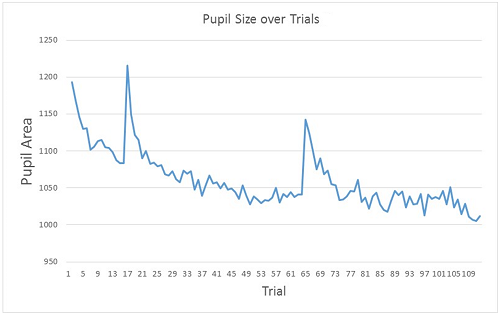
These spikes turn out to coincide with the start of the first and second experimental blocks. The first 16 trials were practice trials. This is a very common pattern and may reflect a range of factors. At the start of the experiment the setting / equipment and task demands are all novel, but as trials progress, participants rapidly learn the task requirements, and general arousal levels reduce (perhaps even boredom sets in…). At the beginning of each block, arousal levels briefly increase as the participant readies themselves for the next effort. The extent to which factors such as fatigue / boredom / learning etc. contribute to patterns such as this is unknown. The important point is that the effect is very robust and, as such, absolute measurements of pupil area may be less meaningful than measurements of change in pupil area within a trial. For this reason, many researchers choose to include a baseline period within each trial, against which subsequent change in pupil area can be measured.
Be Aware of Luminance Changes
Changes in luminance will of course have a profound impact on pupil size. So if you do include a baseline on each trial, it is worth giving some thought as to what should be shown during this period. Having a simple fixation cross could potentially be problematic, particularly if the subsequent trial stimuli are images. The trial stimuli are likely to induce a change in pupil area due to the change in overall luminance levels (rather than any cognitive operations that may have occurred whilst processing the stimuli). The luminance-based change may overshadow any change due to cognitive operations. As such, it can be useful to have an isoluminant baseline. For example, if your study involves presenting different high or low valence words you could have XXXXX as the baseline stimulus, and then BRICK or DEATH as the experimental stimulus. The idea is that you have as many Xs in the baseline as letters in the subsequent experimental stimulus. If your study involves images it can help to use a “scrambled” version of the experimental image as the baseline.
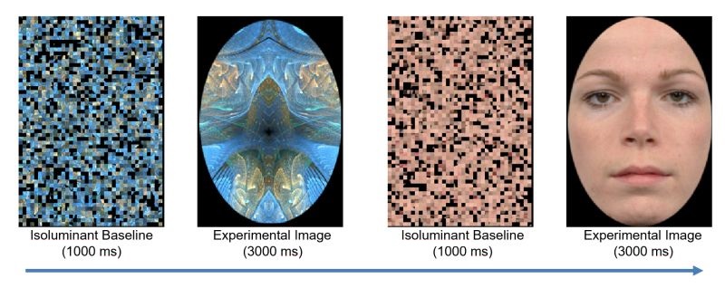
We provide a useful Experiment Builder “helper script” that scrambles the pixels image files on our support forum. Of course, if the appearance of the baseline image itself causes a luminance change (for example, if the previous trial ends with a blank screen, or there was a drift-check between trials), then it is important to leave the baseline image on the screen for long enough for any luminance based pupil size changes to occur.
It can also be a good idea to make sure that the overall luminance levels in the testing room are not too bright or too dark. If the overall environment is too bright then the pupils may not dilate as freely as they would otherwise. If ambient light levels are too dark, then the extent to which the pupil can dilate further may be limited.
The Pupil Foreshortening Effect (PFE) in Pupillometry Research
One potentially important consideration when recording and interpreting pupil area is the pupil foreshortening effect. The camera views the eye from a fixed location. As such, when the eye rotates the pupil appears to change shape (and area). Essentially the pupil appears less circular (and has a smaller area) the further the eye rotates away from the camera lens.

This can result in changes in the reported pupil area of up to 10%. A number of potential solutions have been suggested. Perhaps the simplest is to ensure that all stimuli are relatively small and central – thus minimizing rotations of the eye. Clearly, this is not a viable strategy for tasks in which eye movements are required or expected (such as scene viewing). Another strategy is to ensure that the eye tracker is calibrated and use the gaze data to ensure that there is no systematic difference in gaze location across the experimental manipulation of interest. Finally, there have been attempts to model the PFE, such as the approach outlined by Hayes and Petrov (2016).
Analyzing Pupillometry Data
Simple analyses can be done by calculating a “pupil dilation ratio” or similar measure, by subtracting the average pupil size during the baseline period from the average (or maximum) pupil size during the stimulus period. A more sophisticated approach to pupillometry is to examine the pupillary response itself – for example exploring differences in the duration, onset and even speed of any change in pupil size that occurs in response to stimulus onset.
Our analysis software, Data Viewer, makes both approaches very straightforward. We have lots of useful resources on our support forum (you will need to register to gain access), including a webinar on pupillometry that goes through the steps involved in detail, and instructions for converting pupil area measurements to millimeters.
Final Thoughts
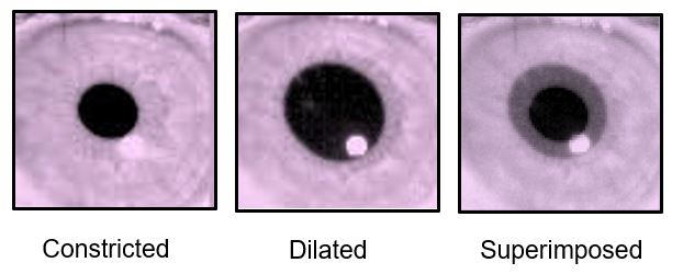
There are many other factors that could be considered in pupillometry research – for example, both caffeine and nicotine can impact on pupil size. Significant individual differences, both in baseline pupil size and pupil motility are common. Another interesting feature of pupillary dilation that often goes unnoticed is that the pupil does not necessarily dilate symmetrically around its center. Nor does it necessarily form a perfect circle, as you can see in the images of my eye below.
There is clearly a great deal more to be learned about the ways in which pupilllometry can reveal insights into cognitive function!
Contact
If you would like us to feature your EyeLink research, have ideas for posts, or have any questions about our hardware and software, please contact us. We are always happy to help. You can call us (+1-613-271-8686) or click the button below to email: